Please use this identifier to cite or link to this item:
https://doi.org/10.21256/zhaw-28264Full metadata record
| DC Field | Value | Language |
|---|---|---|
| dc.contributor.author | Bächinger, David | - |
| dc.contributor.author | Filidoro, Noemi | - |
| dc.contributor.author | Naville, Marc | - |
| dc.contributor.author | Juchler, Norman | - |
| dc.contributor.author | Kurtcuoglu, Vartan | - |
| dc.contributor.author | Nadol, Joseph B. | - |
| dc.contributor.author | Schuknecht, Bernhard | - |
| dc.contributor.author | Kleinjung, Tobias | - |
| dc.contributor.author | Veraguth, Dorothe | - |
| dc.contributor.author | Eckhard, Andreas H. | - |
| dc.date.accessioned | 2023-07-20T13:00:24Z | - |
| dc.date.available | 2023-07-20T13:00:24Z | - |
| dc.date.issued | 2023-06-26 | - |
| dc.identifier.issn | 2045-2322 | de_CH |
| dc.identifier.uri | https://digitalcollection.zhaw.ch/handle/11475/28264 | - |
| dc.description.abstract | We aimed to determine the prevalence of radiological temporal bone features that in previous studies showed only a weak or an inconsistent association with the clinical diagnosis of Meniere's disease (MD), in two groups of MD patients (n = 71) with previously established distinct endolymphatic sac pathologies; i.e. the group MD-dg (ES degeneration) and the group MD-hp (ES hypoplasia). Delayed gadolinium-enhanced MRI and high-resolution CT data were used to determine and compare between and within (affected vs. non-affected side) groups geometric temporal bone features (lengths, widths, contours), air cell tract volume, height of the jugular bulb, sigmoid sinus width, and MRI signal intensity alterations of the ES. Temporal bone features with significant intergroup differences were the retrolabyrinthine bone thickness (1.04 ± 0.69 mm, MD-hp; 3.1 ± 1.9 mm, MD-dg; p < 0.0001); posterior contour tortuosity (mean arch-to-chord ratio 1.019 ± 0.013, MD-hp; 1.096 ± 0.038, MD-dg; p < 0.0001); and the pneumatized volume (1.37 [0.86] cm3, MD-hp; 5.25 [3.45] cm3, MD-dg; p = 0.03). Features with differences between the affected and non-affected sides within the MD-dg group were the sigmoid sinus width (6.5 ± 1.7 mm, affected; 7.6 ± 2.1 mm, non-affected; p = 0.04) and the MRI signal intensity of the endolymphatic sac (median signal intensity, affected vs. unaffected side, 0.59 [IQR 0.31-0.89]). Radiological temporal bone features known to be only weakly or inconsistently associated with the clinical diagnosis MD, are highly prevalent in either of two MD patient groups. These results support the existence of diverse-developmental and degenerative-disease etiologies manifesting with distinct radiological temporal bone abnormalities. | de_CH |
| dc.language.iso | en | de_CH |
| dc.publisher | Nature Publishing Group | de_CH |
| dc.relation.ispartof | Scientific Reports | de_CH |
| dc.rights | https://creativecommons.org/licenses/by/4.0/ | de_CH |
| dc.subject | Human | de_CH |
| dc.subject | Temporal bone | de_CH |
| dc.subject | Radiography | de_CH |
| dc.subject | Magnetic resonance imaging | de_CH |
| dc.subject | Meniere disease | de_CH |
| dc.subject | Endolymphatic sac | de_CH |
| dc.subject.ddc | 617: Chirurgie | de_CH |
| dc.title | Radiological feature heterogeneity supports etiological diversity among patient groups in Meniere's disease | de_CH |
| dc.type | Beitrag in wissenschaftlicher Zeitschrift | de_CH |
| dcterms.type | Text | de_CH |
| zhaw.departement | Life Sciences und Facility Management | de_CH |
| zhaw.organisationalunit | Institut für Computational Life Sciences (ICLS) | de_CH |
| dc.identifier.doi | 10.1038/s41598-023-36479-5 | de_CH |
| dc.identifier.doi | 10.21256/zhaw-28264 | - |
| dc.identifier.pmid | 37365255 | de_CH |
| zhaw.funding.eu | No | de_CH |
| zhaw.issue | 1 | de_CH |
| zhaw.originated.zhaw | Yes | de_CH |
| zhaw.pages.start | 10303 | de_CH |
| zhaw.publication.status | publishedVersion | de_CH |
| zhaw.volume | 13 | de_CH |
| zhaw.publication.review | Peer review (Publikation) | de_CH |
| zhaw.webfeed | Biomedical Simulation | de_CH |
| zhaw.webfeed | Digital Health Lab | de_CH |
| zhaw.author.additional | No | de_CH |
| zhaw.display.portrait | Yes | de_CH |
| Appears in collections: | Publikationen Life Sciences und Facility Management | |
Files in This Item:
| File | Description | Size | Format | |
|---|---|---|---|---|
| 2023_Baechinger-etal_Radiological-feature-heterogeneity-supports-etiological-diversity-Meniers-disease.pdf | Article as published online June 26, 2023 | 6.33 MB | Adobe PDF | 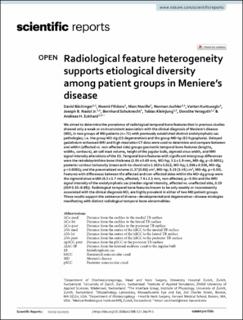 View/Open |
Show simple item record
Bächinger, D., Filidoro, N., Naville, M., Juchler, N., Kurtcuoglu, V., Nadol, J. B., Schuknecht, B., Kleinjung, T., Veraguth, D., & Eckhard, A. H. (2023). Radiological feature heterogeneity supports etiological diversity among patient groups in Meniere’s disease. Scientific Reports, 13(1), 10303. https://doi.org/10.1038/s41598-023-36479-5
Bächinger, D. et al. (2023) ‘Radiological feature heterogeneity supports etiological diversity among patient groups in Meniere’s disease’, Scientific Reports, 13(1), p. 10303. Available at: https://doi.org/10.1038/s41598-023-36479-5.
D. Bächinger et al., “Radiological feature heterogeneity supports etiological diversity among patient groups in Meniere’s disease,” Scientific Reports, vol. 13, no. 1, p. 10303, Jun. 2023, doi: 10.1038/s41598-023-36479-5.
BÄCHINGER, David, Noemi FILIDORO, Marc NAVILLE, Norman JUCHLER, Vartan KURTCUOGLU, Joseph B. NADOL, Bernhard SCHUKNECHT, Tobias KLEINJUNG, Dorothe VERAGUTH und Andreas H. ECKHARD, 2023. Radiological feature heterogeneity supports etiological diversity among patient groups in Meniere’s disease. Scientific Reports. 26 Juni 2023. Bd. 13, Nr. 1, S. 10303. DOI 10.1038/s41598-023-36479-5
Bächinger, David, Noemi Filidoro, Marc Naville, Norman Juchler, Vartan Kurtcuoglu, Joseph B. Nadol, Bernhard Schuknecht, Tobias Kleinjung, Dorothe Veraguth, and Andreas H. Eckhard. 2023. “Radiological Feature Heterogeneity Supports Etiological Diversity among Patient Groups in Meniere’s Disease.” Scientific Reports 13 (1): 10303. https://doi.org/10.1038/s41598-023-36479-5.
Bächinger, David, et al. “Radiological Feature Heterogeneity Supports Etiological Diversity among Patient Groups in Meniere’s Disease.” Scientific Reports, vol. 13, no. 1, June 2023, p. 10303, https://doi.org/10.1038/s41598-023-36479-5.
Items in DSpace are protected by copyright, with all rights reserved, unless otherwise indicated.